
Chairside 26
When the Easier Treatment Plan Threatens the System's Biomechanics
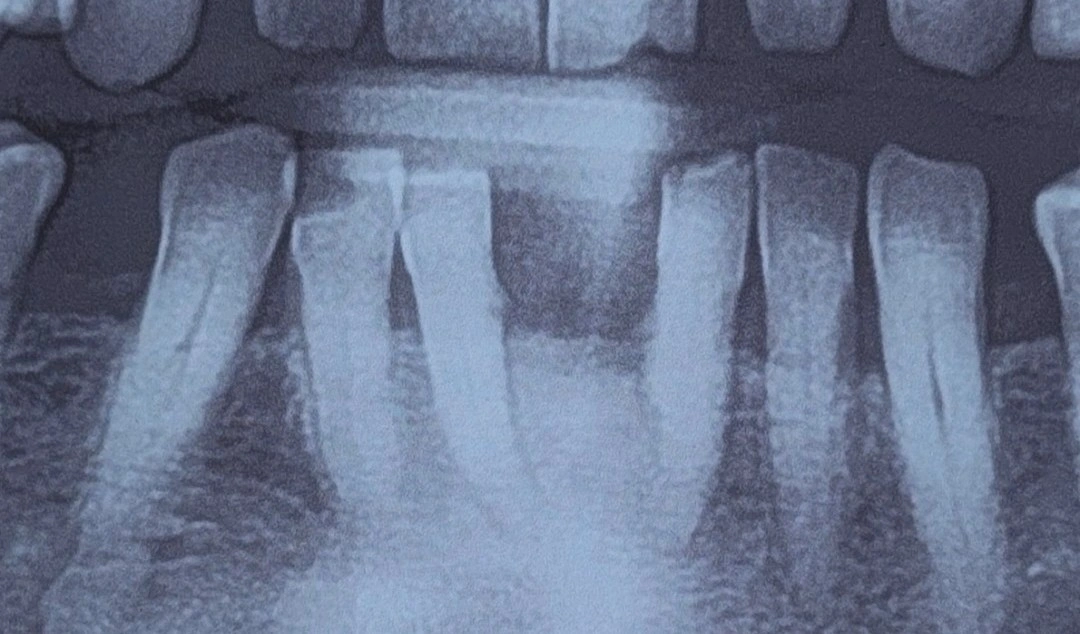
Presenting Complaint. The patient was referred for a specialist prosthodontic evaluation and consultation. The panoramic radiograph showed a large diastema in the anterior mandible, between the two central incisors (teeth #1). Given the poor prognosis, the referring surgeon had judged the central incisors hopeless and extraction-bound; however, because of the need for extensive, complex bone grafting to place an implant in this area, the surgeon's preference was a bridge supported by the lateral incisors (teeth #2) to restore the anterior segment, and a prosthodontic evaluation of the plan was requested.
Clinical and Biomechanical Evaluation. After a careful clinical examination and correlation with the radiographic findings, it was found that the existing gap between the two central incisors was at least as wide as two full teeth. Therefore:
- Evaluating the applied load and span length. If teeth #1 were extracted and teeth #2 were selected as abutments, the resulting bridge would have four pontics in the anterior segment. A span of this length in the anterior mandible is effectively outside the acceptable range for tooth-supported fixed treatment.
- Evaluating the abutments' bone support and root surface. The patient's lateral incisors had a borderline root surface area and bone support. Applying the torque and shear forces generated by four pontics onto two weak abutments conflicts with load-distribution principles and Ante's Law, and made short-term prosthesis failure likely.
- Reviewing the option of extending the abutments to the canines (teeth #3). Had a tooth-supported fixed solution been pursued, the canines would at minimum have had to be brought into the bridge as a long-span design. In that arrangement, the laterals sit between two edentulous spans, and because of their minimal root surface, their contribution to the prosthesis's overall support is negligible — while their presence still constrains the design: preparation of a tooth that adds no meaningful support, difficulty achieving a common path of insertion, an asymmetric distribution of pontics due to the typical position and inclination of these teeth, and harder-to-maintain hygiene in the area. Together, these factors make the cost-benefit ratio of retaining them, in this design, negative.
️Conclusion and Clinical Tip: the proposed treatment plan (a bridge supported by teeth #2) was declined for failing to respect biomechanical balance. The referring surgeon was informed, and the patient was also advised that even if treatment moves toward extracting the lateral incisors and widening the area for an implant, that path is far more logical and standard than needlessly preparing healthy canines and building a long-span bridge with an uncertain prognosis.
This decision, of course, is not absolute — it is case-dependent. If the laterals have a healthy periodontium and a good prognosis, removing a healthy tooth merely to simplify the design conflicts with the principle of minimal intervention. But when their support is borderline, keeping a tooth that adds no support yet constrains the design is not justified.
In prosthodontic planning, an unreasonable biomechanical load should never be shifted onto weak dental abutments simply to avoid surgical challenges such as bone grafting. Anterior treatment shortcuts usually lead to more catastrophic failures.
The content of this page is intended for the educational use of dentists and dental students.