
Insight 73 — Crown Replacement and Soft Tissue Management with Inflammation from Food Impaction
Published:
Last reviewed:
فارسی
Clinical Explanation
This note follows a crown replacement in which two independent decisions were taken side by side: what had to be corrected in the previous preparation, and how the inflamed soft tissue was to be managed before scanning. The patient had presented because of food impaction and the inflammation it had caused, but refining the preparation was not the answer to that complaint and stood on its own. The decision about the retraction cord is likewise taken region by region here, rather than as one uniform protocol around the whole tooth.
-
The patient's complaint
This tooth had a previous crown. The patient's main complaint was food impaction, which had led to pain and gingival inflammation. The cause of the food impaction was a defective proximal contact on the previous crown. -
Crown removal and preparation refinement
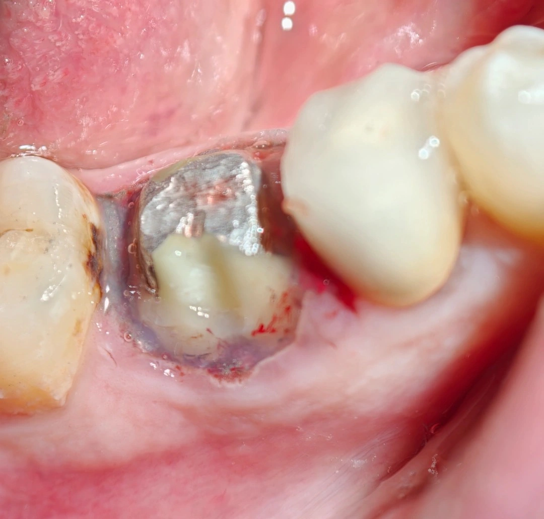
The crown was removed and the preparation was refined first. One point needs to be made clear: refining a preparation is not necessarily related to the patient's subjective complaint. The preparation was refined here because the previous preparation was defective, not because the patient was complaining of food impaction. The defects of the previous preparation: the amalgam had an overhang on both sides, the finish line was inadequate, and the walls needed correction. The finish line and the walls were corrected. The contact problem, which was the cause of the patient's complaint, is resolved at the design stage of the new crown. -
Gingival condition after preparation refinement
After the preparation was refined, the gingiva was bleeding. Before deciding about retraction, the circumference of the tooth was assessed. The only area with a sulcus that could be retracted was the lingual side. The retraction cord was placed in that area only. On the proximal surfaces and the buccal, the tissue was completely attached to the tooth and there was no retractable sulcus. Placing a cord in these areas would only have caused more trauma and more bleeding, so no cord was placed there. -
Bleeding control
To control the bleeding, an aluminium-based hemostatic gel was placed around the tooth and two minutes were allowed to pass. After fluid control and drying of the area, the scan was taken. -
Clinical point
Cord retraction is not a fixed protocol for every area. The decision to place a cord has to be made on the basis of the actual condition of the sulcus in each area. Where there is no retractable sulcus, a cord only adds damage and makes the quality of the scan worse.
The content of this page is intended for the educational use of dentists and dental students.