
Save the Tooth or Extract It? Three Alternative Treatments from a Prosthodontic Perspective
Surgical Extrusion, Intentional Replantation and Tooth Autotransplantation
Introduction: Where Does the Treatment Decision Begin?
When the crown of a tooth is severely destroyed, a fracture has reached below the gingiva, or a previous endodontic treatment has failed, the decision between saving the tooth and extracting it is not always simple. From a prosthodontic perspective, one fundamental question has to be asked before choosing a treatment: if the tooth is saved, can a reliable restoration with a good prognosis ultimately be made for it?
A root may be treatable endodontically or surgically, yet the remaining structure, the bone support or the occlusal conditions may not allow an acceptable restoration. Conversely, a tooth that looks unrestorable in its current position can sometimes become restorable through a change of position or an appropriate surgical intervention.
The three treatments discussed in this article make it possible, in selected patients, to keep the natural tooth or to replace it with another natural tooth:
- Surgical Extrusion: moving the root to a more coronal position within the same socket, so that an unrestorable tooth becomes restorable.
- Intentional Replantation: deliberately extracting the tooth, treating the root problem outside the mouth, and returning it to its original position.
- Tooth Autotransplantation: moving an impacted or erupted tooth from one area of the mouth to another area in the same patient.
The protocols of these three treatments are very similar: atraumatic extraction of the tooth, direct inspection of the root, and its replacement in the socket. That is why, in all three, the success of treatment depends on preserving the health of the periodontal ligament (PDL).
1. Surgical Extrusion: When the Main Problem Is Restorability
Suppose a patient presents with a crown-root fracture of a maxillary central incisor. On the labial side the fracture margin is close to the gingiva, but on the palatal side it extends below the bone. In such a situation the root may be maintainable endodontically, but there is not enough ferrule for the final restoration.
One option is surgical extrusion. In this technique the root is moved with minimal damage and placed in a more coronal position, so that more sound structure is available for the restoration. So the goal is not just to bring part of the root out; the goal is to make room for a reliable restoration.
Case Selection from a Prosthodontic Perspective
The main indication for surgical extrusion is single-rooted permanent teeth with a crown fracture or caries near the bone level, and the patient's age is not a limitation. Before deciding, it must be clear what conditions we will actually have after the root has been repositioned.
Remaining structure and ferrule: before treatment, all carious dentine must be removed so that the real sound structure can be seen and the amount of extrusion needed can be determined. If an adequate ferrule cannot be obtained even after extrusion, the treatment has not solved the main problem.
Root length and bone support: with extrusion, part of the root that was inside the bone moves coronally. The root must therefore be long enough after extrusion to have sufficient alveolar support and an appropriate crown-to-root ratio. Creating a ferrule should not come at the cost of losing the root's support.
Even so, in young patients extruding even a short root may count as a success, provided that temporarily keeping the tooth can postpone an implant until adulthood.
180-Degree Rotation in Maxillary Anterior Teeth
In crown-root fractures of maxillary anterior teeth, the fracture line follows a characteristic pattern: on the labial side it is at or above the gingival level, and on the palatal side it often runs below the bone. At the same time, in this region the alveolar bone level on the palatal side is more coronal than on the labial side.
For this reason, rotating the root by 180 degrees before placing it back in the socket is an ideal technique for these teeth. With this rotation, the deep palatal margin moves to the labial side, where the bone is lower, and the shallow labial margin moves to the palatal side. The result is that the fracture margin on both sides becomes accessible with the least amount of extrusion, and more of the root length stays inside the bone. In maxillary premolars, 180-degree rotation does not seem to affect the treatment outcome.
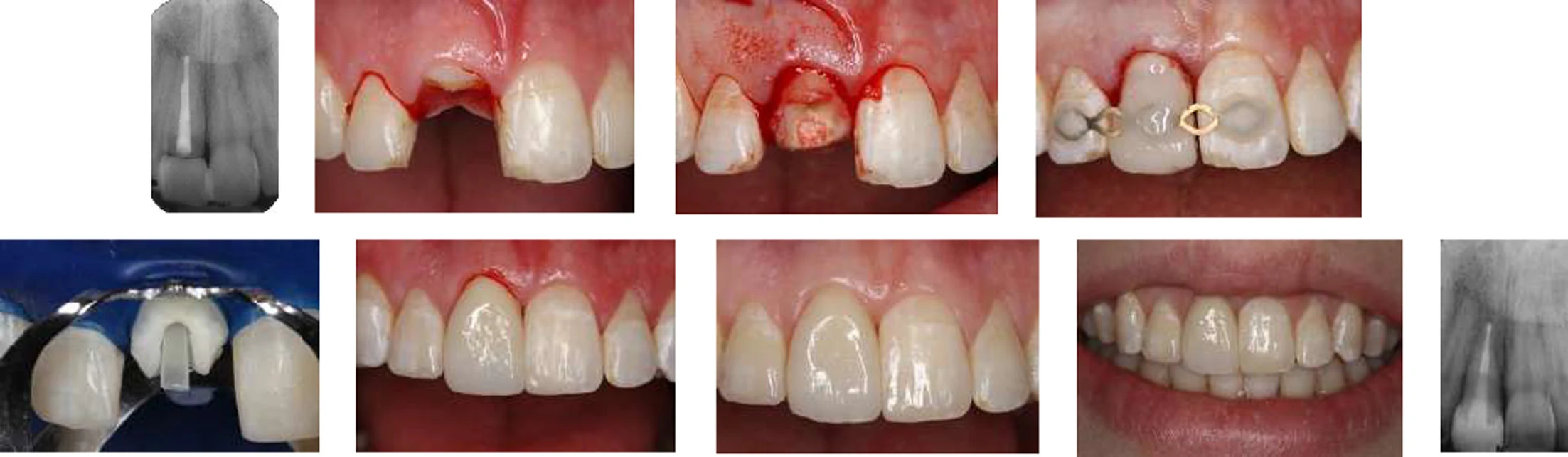
Surgical Extrusion or Crown Lengthening?
In surgical crown lengthening, bone usually has to be removed to reach sound tooth structure. This can affect aesthetics, the gingival margin and the support of the adjacent teeth, especially in the anterior region. In surgical extrusion, instead of removing bone, the root itself is moved. Even so, none of these techniques is the best choice for every patient, and the choice between surgical extrusion, orthodontic extrusion (forced eruption) and crown lengthening should be made by weighing the benefits and risks of each technique.
Splinting, Root Canal Treatment and the Timing of Restorative Treatment
After surgical extrusion, the tooth is stabilised with a flexible splint, for example a flexible wire bonded to the adjacent teeth with composite. The aim is not to immobilise the tooth completely; the splint should provide enough stability while allowing physiological movement, so that the risk of ankylosis is reduced. Because the extruded root does not fit the socket, the splinting period may be longer than for avulsed teeth and reach up to six weeks, especially when root mobility is high.
Following the protocol for avulsed teeth, root canal treatment should begin within the first two weeks after the root is placed back in the socket, to prevent infection-related root resorption. In the first week, calcium hydroxide is not recommended as an intracanal medicament, because it may have a negative effect on periodontal healing.
Restorative treatment is usually carried out six to eight weeks after extrusion. If the tooth needs a crown, preparation with BOPT (Biologically Oriented Preparation Technique) can help achieve a better gingival form and longer-term soft tissue stability. Before the final restoration, the stability of the tooth, the condition of the soft tissue and the health of the periodontium should be reassessed.
What Counts as Success?
Surgical extrusion is successful when periodontal healing occurs without root resorption or ankylosis, the marginal bone level remains stable, and tooth mobility does not increase significantly. In patients whose growth is complete, ankylosis can also be an acceptable outcome, because replacement resorption progresses slowly in them and does not cause significant infraposition.
2. Intentional Replantation
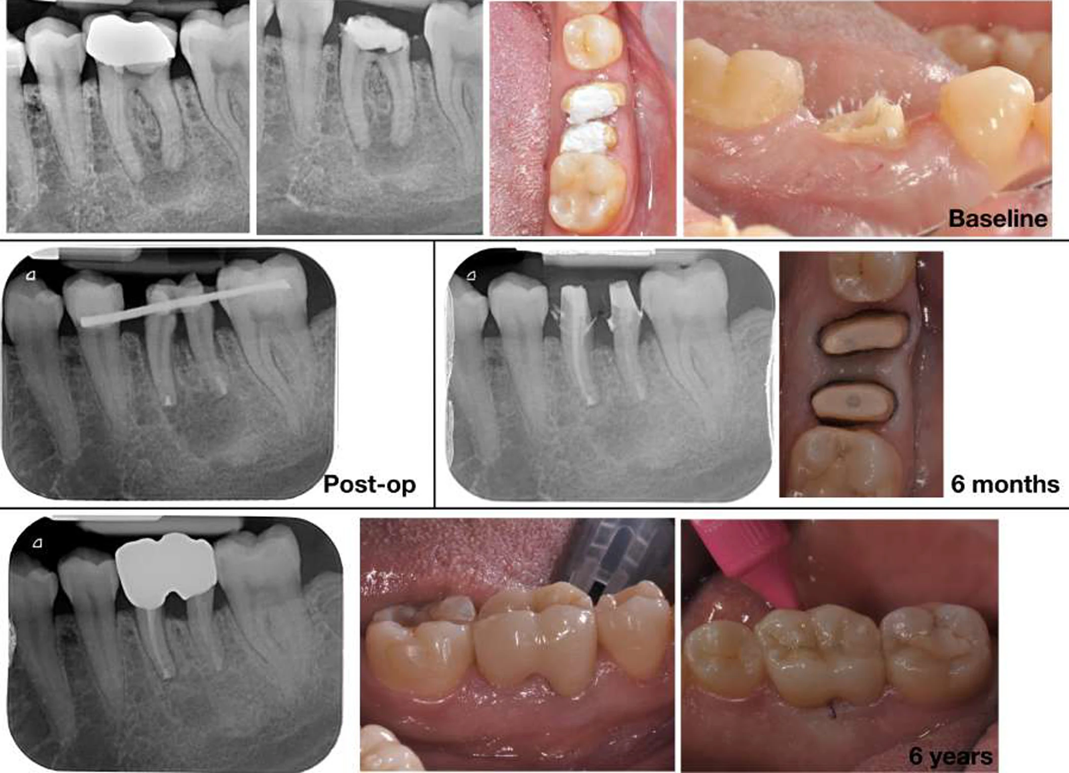
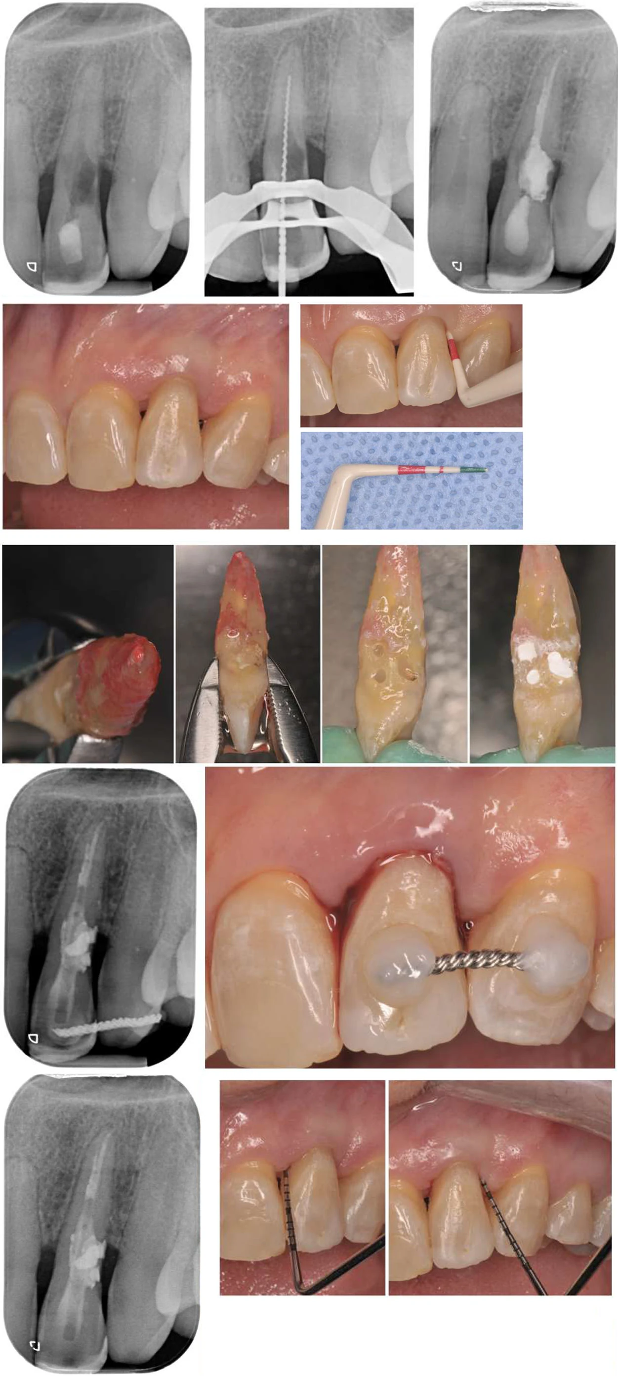
In intentional replantation, the tooth is deliberately extracted so that a problem that is hard to reach can be treated outside the mouth, and it is then returned to the socket at the same bone level as before; that is, unlike surgical extrusion, the position of the tooth does not change. Its main indication is failed endodontic treatment when retreatment or apical surgery is not possible, for example because the root is close to the mental nerve or the maxillary sinus. Inaccessible perforations and external cervical resorption are other indications (Figure 3). This technique can also be combined with surgical extrusion, as in the case in Figure 2, where a molar without ferrule was treated with replantation, surgical extrusion and root separation.
In a systematic review, the survival of replanted teeth was reported at about 90 percent. The most important prognostic factor is extraoral time, and when this time exceeded 15 minutes survival was lower; the root surface must also stay moist throughout this period. From a prosthodontic perspective, this treatment only makes sense when the root problem is the only obstacle to keeping the tooth and the coronal structure is sufficient for a restoration; a lesion being treatable is not the same as the tooth being restorable. Treating vertical root fractures with this technique is still not acceptable, because its long-term results have been unfavourable.
3. Tooth Autotransplantation
In tooth autotransplantation, a tooth is moved from one area of the mouth to another area in the same patient, for example a third molar to the site of a second molar that cannot be kept. Its main advantage over an implant is the presence of a PDL: the transplanted tooth can move along with growth and the eruption of the adjacent teeth, whereas an implant stays fixed in a growing patient. For this reason it is a valuable option in young patients, but it also has applications in adults, and in a systematic review the five-year survival of teeth with complete root formation was reported at 90.5 percent. An immature tooth may revascularise, but a tooth with a complete root usually needs root canal treatment.
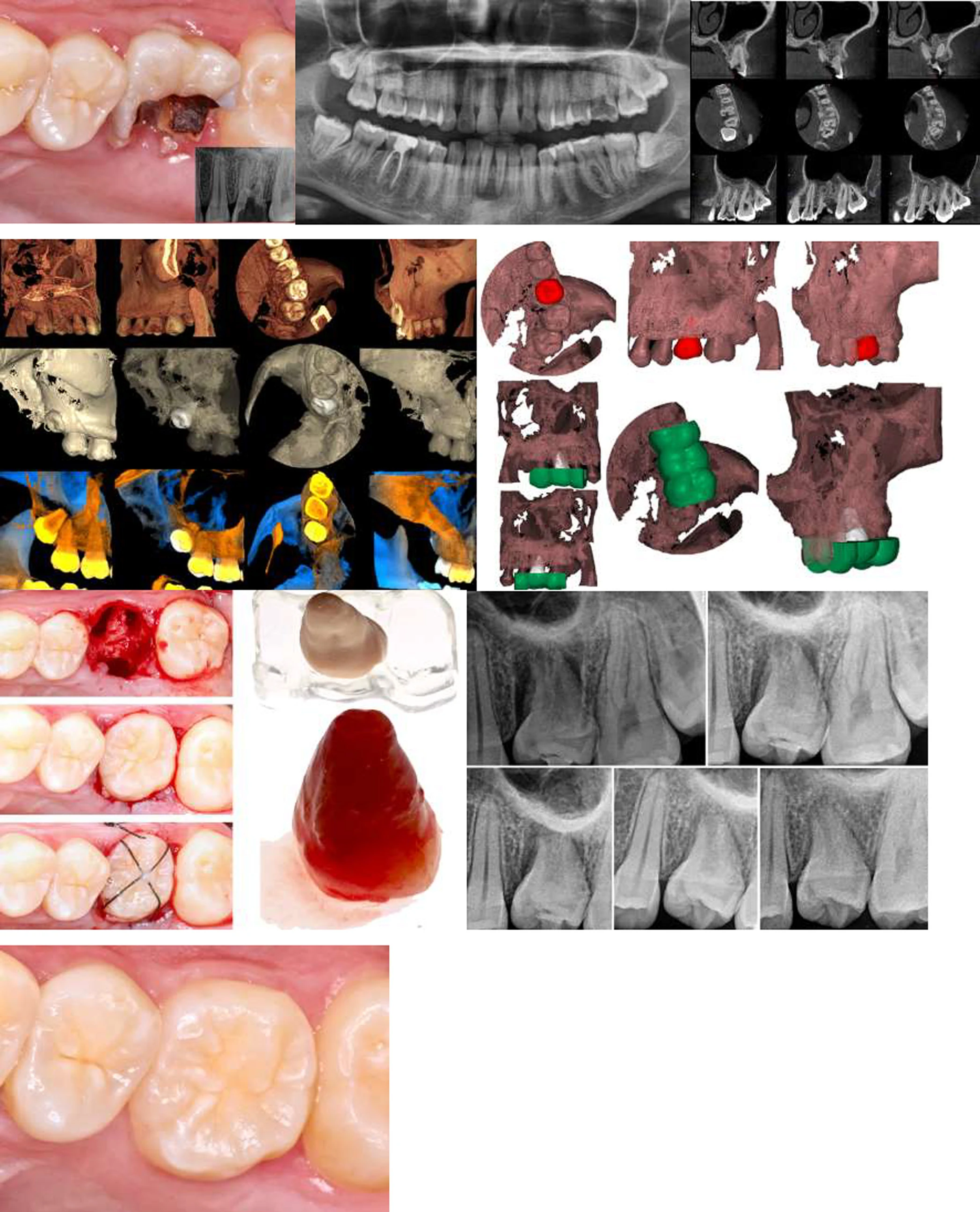
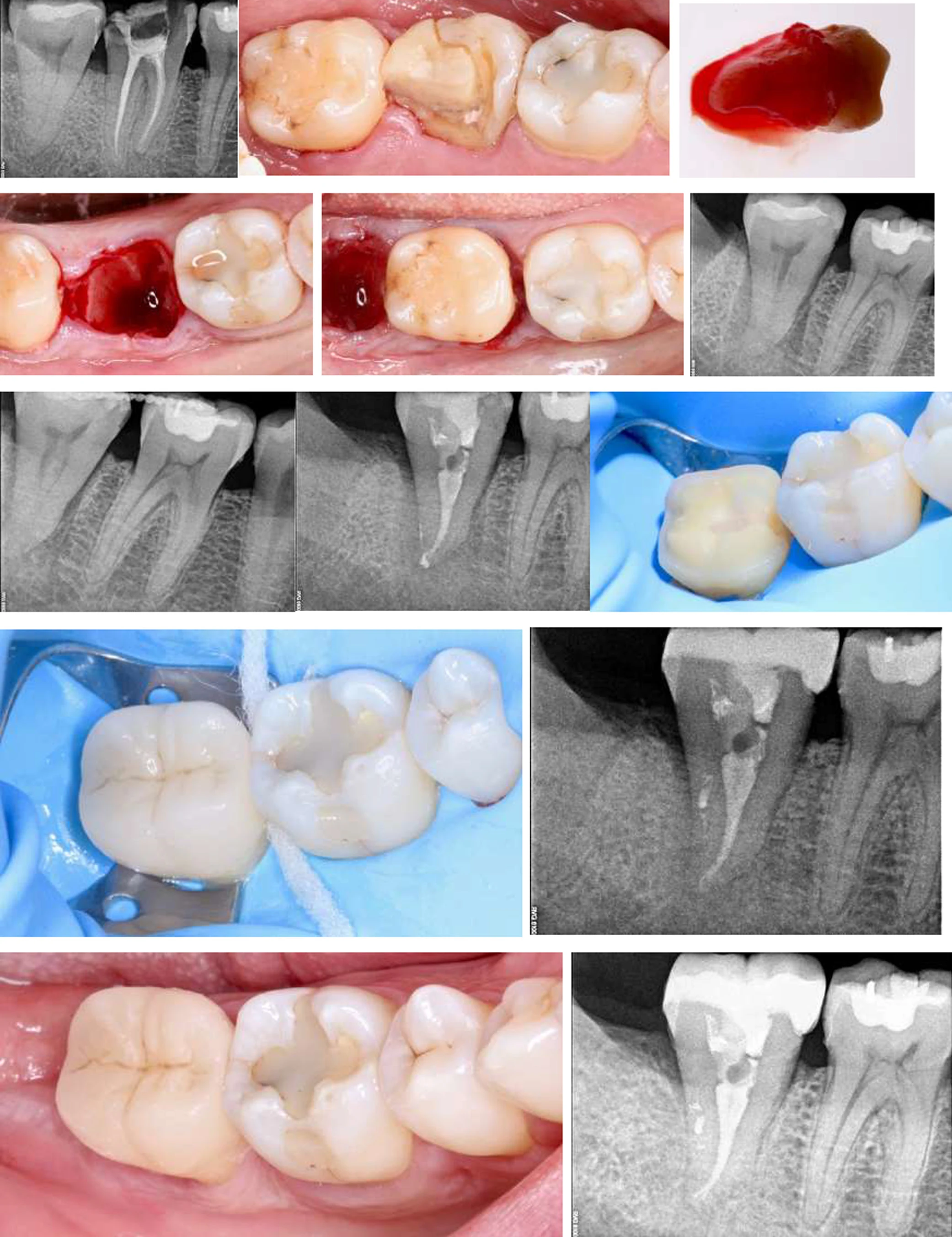
From a prosthodontic perspective, the donor tooth must match the recipient space in terms of crown dimensions, final position and the possibility of building an acceptable occlusion. Planning with CBCT and a 3D replica helps in selecting the tooth and preparing the socket more precisely, and reduces extraoral time (Figure 4). The tooth is placed slightly below the occlusal plane, stabilised with sutures or a flexible splint, and restorative treatment is carried out after healing. In the case in Figure 5, the transplanted third molar was restored with a lithium disilicate overlay.
4. Why Is PDL Health So Important?
In all three treatments, success depends on the survival of the PDL cells. Mechanical damage to the root surface during extraction, drying of the root surface, or prolonged extraoral time increases the likelihood of external root resorption and ankylosis.
Ankylosis is a more serious problem in a growing patient, because the ankylosed tooth does not move with bone growth and the eruption of the adjacent teeth, goes into infraposition, and also arrests the growth of the alveolar bone. In a patient whose growth is complete, ankylosis can be an acceptable outcome, but it still does not mean normal periodontal healing. For this reason, seating the tooth in the socket is not the end of treatment, and clinical and radiographic follow-up is necessary.
5. Limitations of the Scientific Evidence
These three techniques have been used clinically for decades, but the level of evidence supporting them is still limited. Many studies have small sample sizes and lack adequate long-term follow-up. At the time the source of this article was published (2022), there was only one animal study comparing surgical extrusion with orthodontic extrusion, and comparative clinical studies are needed. The role of planning with CBCT and 3D replicas, the effect of orthodontic forces on extruded or replanted teeth, and newer approaches such as cryopreservation of donor teeth and the use of growth factors also still need further investigation.
The reported survival percentages should also not be generalised to every case without considering patient selection, the initial condition of the tooth and the length of follow-up.
Conclusion: Saving a Tooth Should Serve the Treatment Plan
Surgical extrusion, intentional replantation and tooth autotransplantation show that for some severely damaged teeth there are options other than extraction and replacement with an implant. But the choice of surgical extrusion should not be based only on whether the surgery or the endodontic treatment can be done.
From a prosthodontic perspective, it is better to define the final outcome first: can we reach a tooth with adequate periodontal support, sufficient structure, an acceptable occlusion and a reliable restoration? Then the endodontic, periodontal, orthodontic or surgical treatments should be chosen to reach that same goal. The tooth is not meant to be saved at any cost; a treatment is worth doing when it has an acceptable biological, mechanical and functional prognosis.