
How Can We Replace an Anterior Tooth Temporarily After Implant Placement?
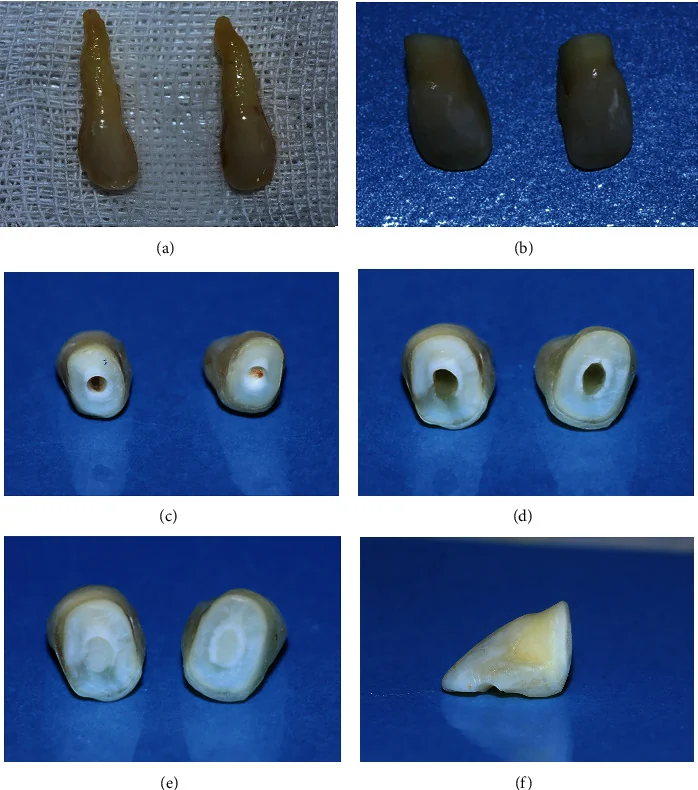
1. Using the Patient’s Natural Crown as a Pontic
When the implant is placed at the time of extraction and the natural crown is intact, the crown can sometimes be reused as a pontic.
The root is removed, the fitting surface of the crown is adjusted to suit the soft tissue and polished, and the crown is attached to the adjacent teeth with composite or fiber reinforcement.
The main advantage is that the patient’s own tooth preserves its natural shade, shape, translucency, and anatomy. This can be valuable in the anterior region, where matching color and form matters.
The crown must have enough sound structure, and it must be possible to attach it without creating an unfavorable occlusal contact.
2. A Composite Pontic or a Prefabricated Tooth
If the patient’s natural crown cannot be reused, the pontic can be built directly with composite or made from a prefabricated acrylic or composite tooth.
The artificial tooth is adjusted for size, shade, and shape, then bonded to the adjacent teeth with resin. Alternatively, a pontic can be shaped directly in the mouth with composite.
These approaches are relatively quick and inexpensive, and they preserve the adjacent teeth.
Their main limitation is the strength of the bond. Debonding or fracture is more likely in patients with a deep bite or heavy anterior contacts.
3. A Fiber-Reinforced Composite Bridge
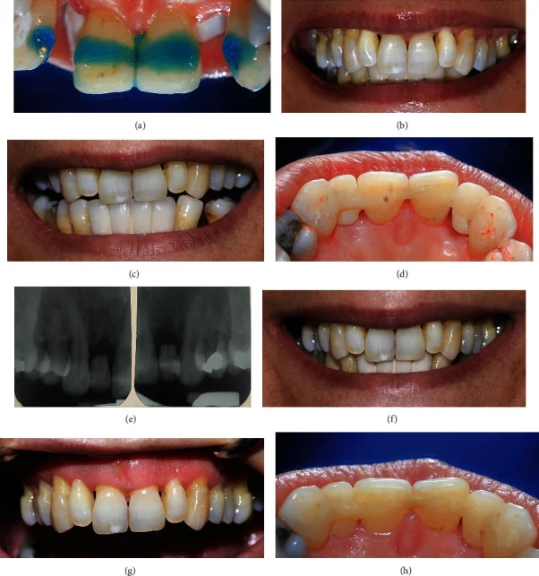
In a fiber-reinforced composite (FRC) bridge, fiber reinforces the bridge and helps attach the pontic to the adjacent teeth.
The pontic itself may be made from composite, a prefabricated tooth, or even the patient’s natural crown. FRC describes the reinforcement and connection approach; it does not mean there is only one type of pontic.
The bridge can be made directly at chairside or indirectly.
It offers a fixed replacement that does not rely on the implant and usually requires little or no preparation of the adjacent teeth. If it fractures or debonds, repair may be possible in many cases.
Success depends on having enough bonding surface, enough room for the fiber, and control of occlusal forces.
4. A Temporary Resin-Bonded Bridge
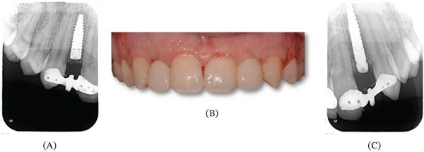
Another option is a temporary bridge with resin-bonded retainers, similar to a Maryland bridge.
It may have one or two retainers bonded to the palatal surfaces of the abutment teeth. In a single-retainer design, the pontic is attached to only one tooth, so independent movement between two abutment teeth is less likely to challenge the bond.
This is a fixed option that usually requires little preparation of the adjacent teeth. It may be useful when the waiting period is expected to be longer.
Adequate enamel for bonding and enough space for the retainer are important. A deep bite or heavy palatal contacts can make the design and placement difficult.
5. An Essix Retainer with a Pontic
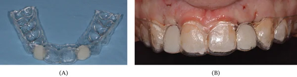
An Essix retainer is a clear appliance made to fit over the teeth. The missing tooth space is filled with a pontic or tooth-colored material.
It is relatively simple and inexpensive to make, and it does not require preparation or bonding of the adjacent teeth. With a suitable design, contact with the surgical site can be reduced.
It is removable, so patients usually take it out for meals. Covering the occlusal surfaces may also feel uncomfortable to some patients.
This option is useful when we want to avoid placing any resin-bonded attachment on the adjacent teeth.
6. An Acrylic Flipper
An acrylic flipper is a common removable option for temporarily replacing an anterior tooth. It is usually made of acrylic and rests on nearby teeth and tissues to fill the space.
It is relatively simple and inexpensive to make, and it can be adjusted during healing.
After recent implant placement, the fitting surface needs particular attention. Unwanted contact or pressure on the surgical site, healing abutment, or graft may interfere with healing.
Retention and stability also matter. A small, unilateral flipper that dislodges may pose a swallowing or aspiration risk.
Its design should not be based only on appearance or on making the appliance as small as possible.

How Do We Choose?
The choice depends on the adjacent teeth and available bonding surfaces, the patient’s occlusion, soft-tissue conditions, whether grafting was performed, the expected time before the definitive crown, and whether the patient prefers a fixed or removable option.
If the natural crown is intact, it may be an attractive pontic. When bonding is suitable, an FRC or resin-bonded bridge can provide a fixed, conservative option. An Essix retainer or flipper may be useful when a removable replacement is preferred.
Across all these options, the shared goal is to fill the space and maintain appearance without applying harmful pressure to the implant or healing tissues.
Replacing the missing tooth does not necessarily create an appropriate emergence profile. Depending on the soft-tissue condition, an implant-supported provisional crown may still be needed after osseointegration to shape the gingival contour.