
RPD Design in the Digital Era: What Has Really Changed?
Suppose a patient with bilateral free-end edentulism in the mandible has come to us for a removable partial denture. We can scan the patient's mouth, build a digital model and even produce the metal framework by 3D printing. But who is going to determine the path of insertion, the location of suitable undercuts, the position of the rests and the design of the clasps?
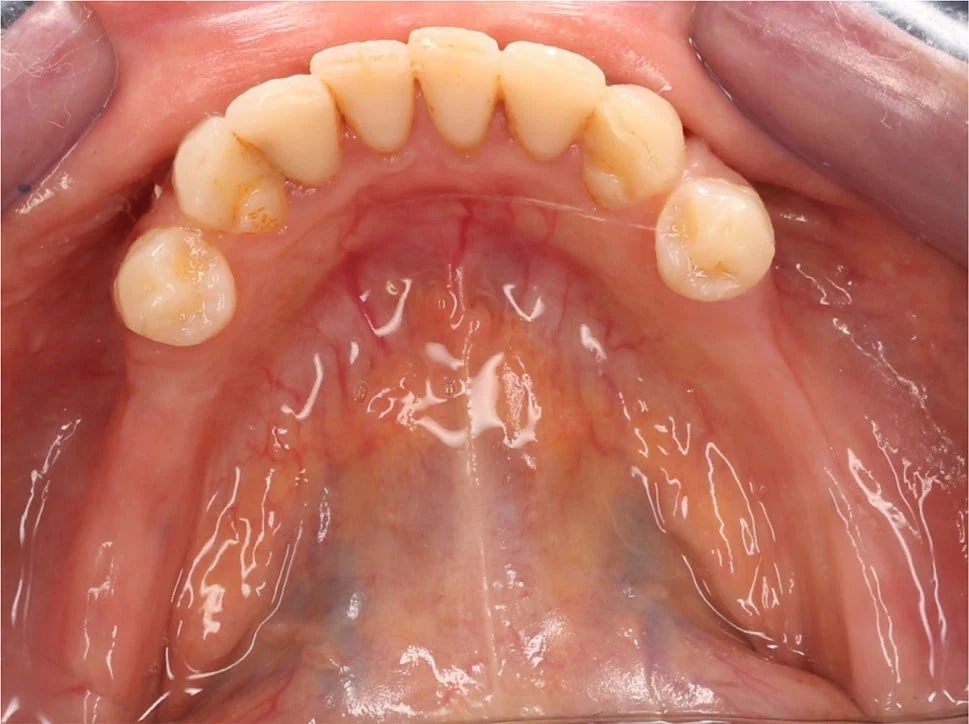
In fact, digitizing the fabrication steps does not mean that the design of the prosthesis has been done automatically.
To understand this, it helps to separate three stages: recording the information of the mouth, designing the prosthesis and fabricating it. Each of these stages can be carried out by a conventional or a digital method.
For example, we might take a conventional impression, scan the stone cast and continue the design and fabrication digitally. Or we might have an intraoral scan from the start, but still do the initial design by hand. This distinction is also seen in the 2026 systematic review by Alqutaibi et al., where conventional, semi-digital and digital workflows are separated from one another.
But RPD design deserves special attention. Because a 3D model records only the shape of the teeth and tissues, whereas the design must specify how we use those structures to provide support, retention and stability for the prosthesis.
One of the first decisions is the choice of the path of insertion.
In the conventional method, we place the cast on the surveyor and examine different paths of insertion by changing its tilt. Then we mark the line of greatest convexity of the tooth, the height of contour, and the usable undercuts.
The position of the usable undercuts depends on the path of insertion. By changing the tilt of the cast, we may find a more suitable undercut on one tooth, but that same change may make the guide planes or the undercuts of other teeth unsuitable.
So surveying is not just a geometric measurement; it is part of prosthetic decision-making.
On the other hand, RPD design is not limited to choosing a clasp. The rests must provide adequate support, the guide planes must direct the path of insertion and removal, and the various components of the prosthesis must be coordinated in a single design. Therefore, identifying one undercut is not enough for proper RPD design.
Now let us see what digital technology has changed.
In digital surveying, instead of a physical cast we examine the 3D model in software. We can change the orientation of the model, evaluate different paths of insertion, and see the survey lines and undercuts on the model.
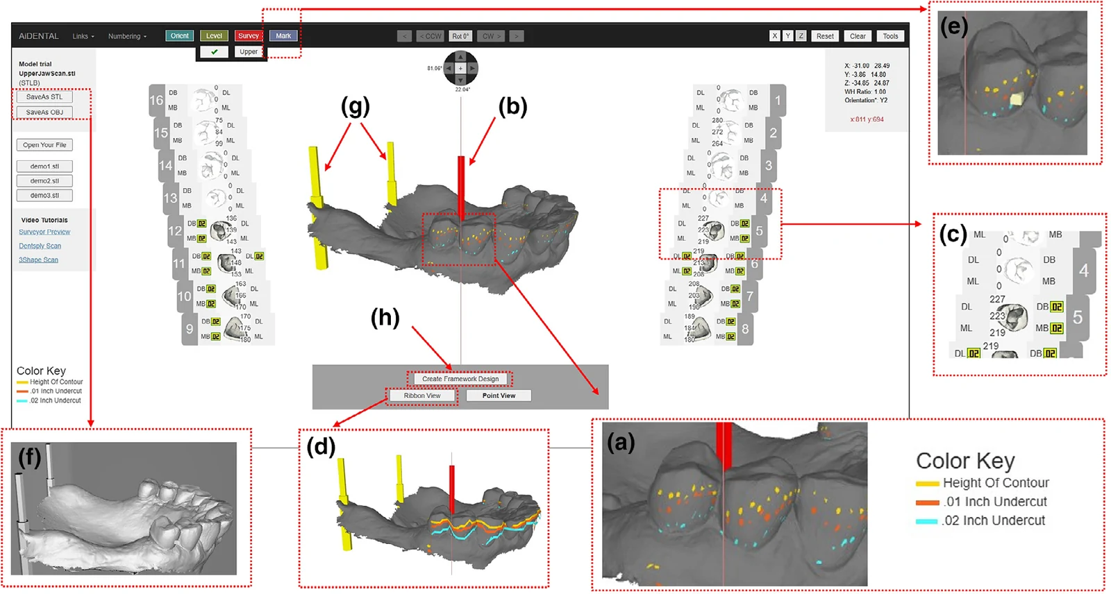
But perhaps more important than the digital display of undercuts is the ability to record and retrieve the survey information.
In the conventional method, once we have chosen a suitable path of insertion, we must be able to put the cast back in the same position; this is one of the purposes of tripoding.
In the digital environment, the selected path, the position of the undercuts and the design markings can be saved with the model so that they can be retrieved and reviewed later.
This capability becomes important in communication between the dentist and the laboratory.
If we send only the patient's scan file to the laboratory, the information about the design decisions has not necessarily been transferred with it. As a result, the laboratory may have to repeat the survey and part of the design.
But in an integrated digital workflow, the survey information and the prosthesis design can be transferred together with the model. In effect, instead of sending merely the shape of the teeth, we also send the design decisions to the laboratory.
Mahrous et al., in a 2026 article, drew attention to this very gap between the digital recording of information and digital RPD design.
They introduced a web-based system that makes it possible to select and save the path of insertion, identify undercuts and prepare a proposed RPD design. The dentist can modify components such as the clasps, rests and major connector, and transfer the design information to the laboratory. Even reconstructing the selected position on a physical surveyor is envisaged.
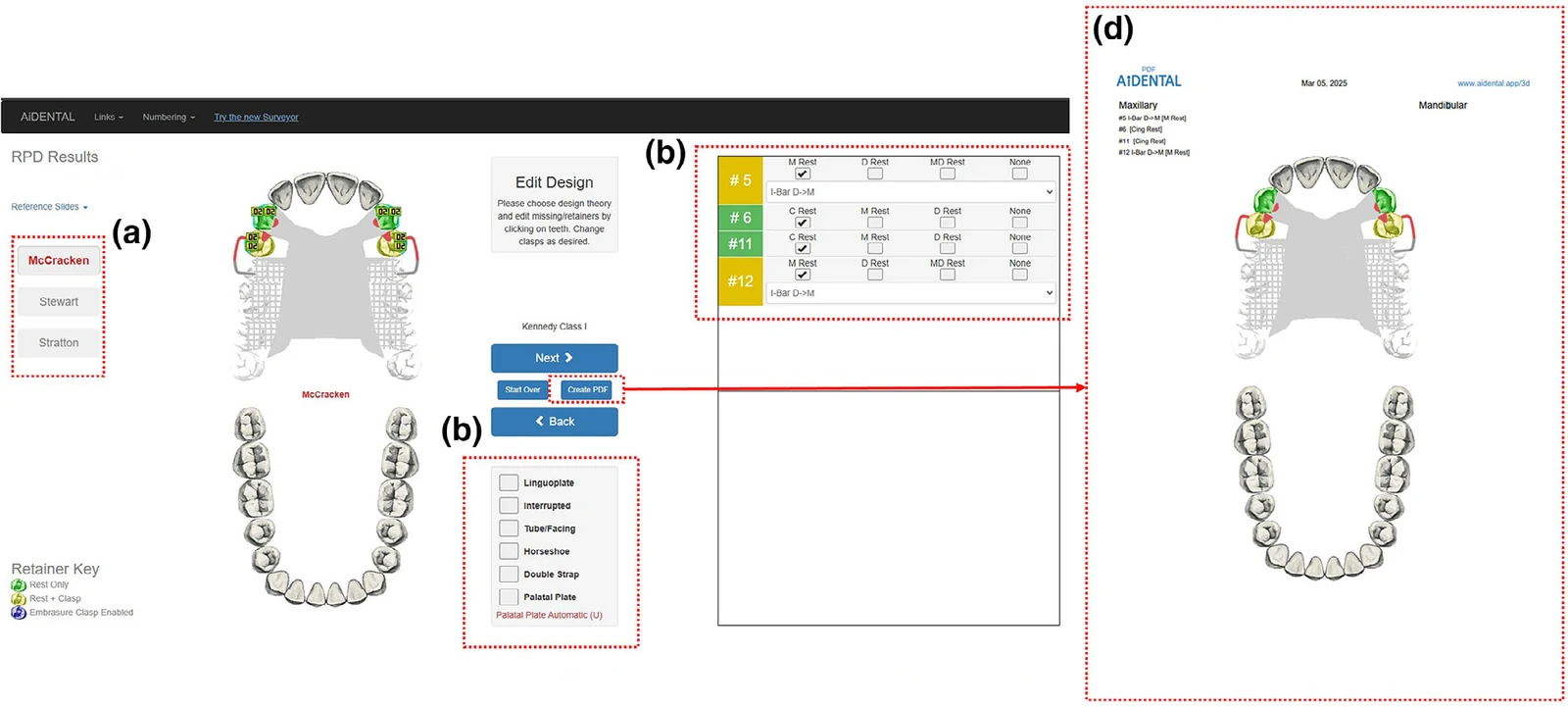
What matters about this article for us is not the name of the software; it shows how diagnostic surveying and initial design can be connected to the digital workflow.
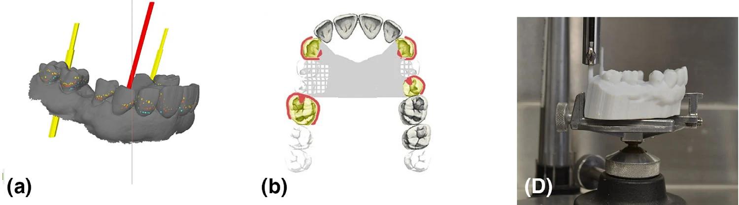
Of course, another question arises here: when software can identify undercuts and even offer a proposed design, can prosthetic decision-making be entrusted to it as well?
We have to distinguish between geometric calculation and clinical judgment.
Software can calculate the position of undercuts or propose a design based on specified rules. But whether that design is suitable for the patient depends on information beyond the geometry of the model, including the condition of the abutment teeth, periodontal status, the way the prosthesis is supported and occlusal considerations.
The authors of the article also stress that the software's suggestions should not replace the dentist's clinical judgment.
On the other hand, if the scan is incomplete, has artifacts or the teeth are malpositioned, automatic detection of undercuts may be erroneous. So a calculation being digital does not mean its result is definitely correct.
But what does the evidence say about the benefits of this method?
The article by Mahrous et al. is a technique-description article, not a study that has compared patient outcomes with the conventional method.
The authors put forward advantages such as saving time, reducing manual steps and improving communication with the laboratory, but they provide no comparative data to prove these advantages.
Therefore, it is still not possible to conclude that using such software necessarily leads to better framework fit, greater retention or better long-term RPD success.
Another point is the independence of the different stages of a digital workflow.
For example, Elgamal et al., in a clinical study, examined two routes for obtaining the digital model: one with a conventional impression and cast scanning, and the other with direct intraoral scanning. Nevertheless, the framework design was the same in both routes.
This example shows that digital recording of information and digital design are two independent stages, and the effect of a change in each stage can be examined separately.
Overall, the main change in digital RPD design is not merely the replacement of the physical surveyor with software; it is the ability to record, modify, retrieve and transfer design decisions in a digital environment.
Nevertheless, the principles of design have not changed. We still have to choose a suitable path of insertion, consider support and retention together, and design the components of the prosthesis according to the patient's conditions.
Digital technology has provided new tools for making and recording these decisions, but the correctness of the decisions still depends on the dentist's knowledge and judgment.
∆ References
- Systematic review of 3D-printed RPD metal frameworks compared with conventional and partially digital methods (Alqutaibi et al., 2026)
- A novel digital diagnostic surveyor and designer for removable partial dentures (Mahrous et al., 2026)
- Cross-over clinical study of the accuracy of SLM RPD frameworks made with two workflows (Elgamal et al., 2025)