
Digital Full-Arch Implant Scanning: How Far Can We Really Trust It?
Suppose we have a patient with four or six implants in an edentulous jaw, and we are going to make a fixed prosthesis for them.
To make this prosthesis, the positions of the implants relative to each other have to be recorded accurately. If the distance or angulation between them is not recorded correctly, the final prosthesis may not fit properly.
So the question is: can an intraoral scanner do this as accurately as a conventional impression?
A new systematic review in JPD, examining 23 studies, compared different digital scanning methods with the conventional open tray impression.
The reference method in these studies was an open tray impression with impression copings splinted together at the abutment level; that is, we tighten the impression copings onto the multi-unit abutments, splint them together with a material such as pattern resin, and then take the impression.
What Is the Main Problem with Full-Arch Scanning?
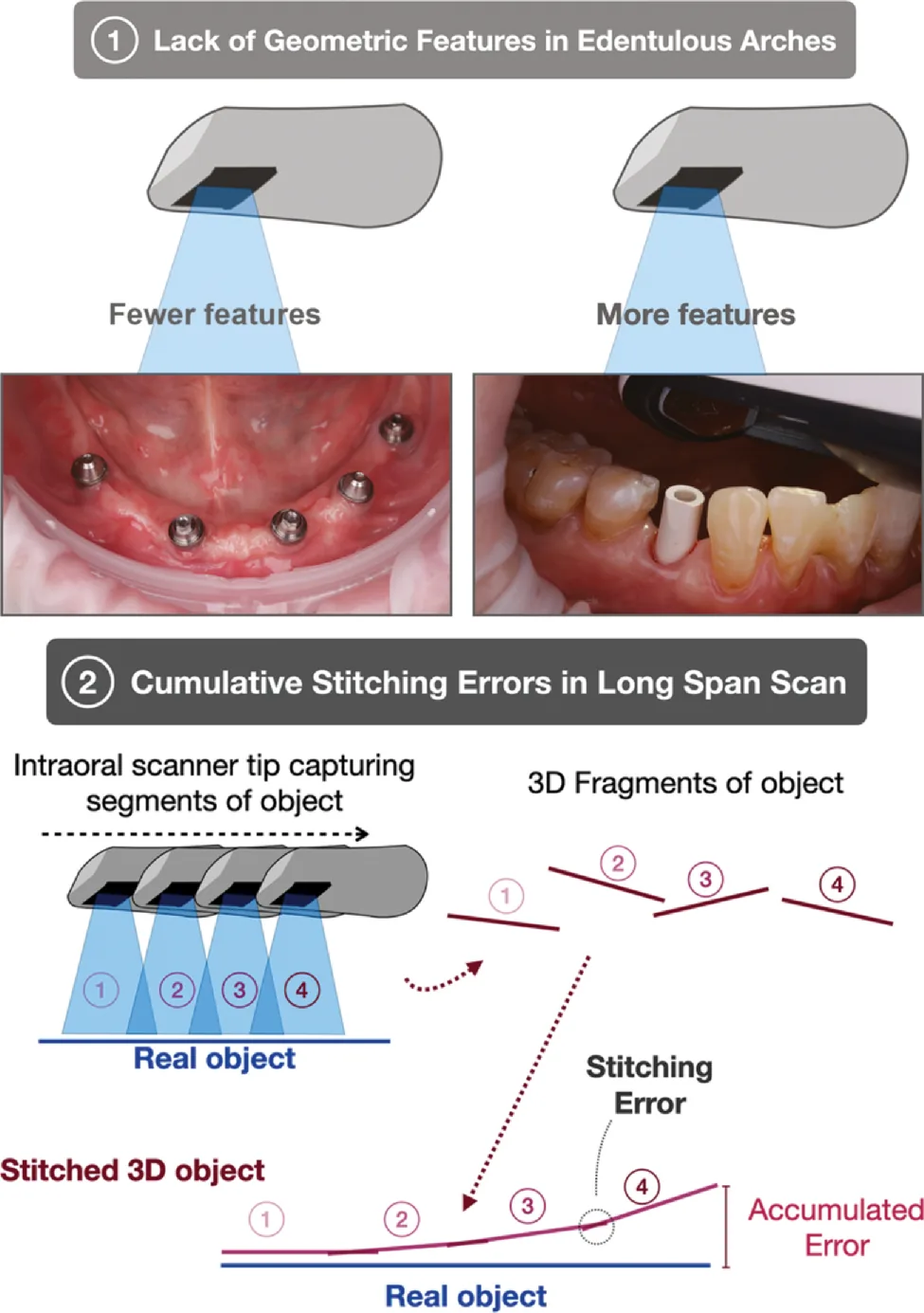
When scanning natural teeth, cusps, fissures and tooth shape help the software recognize its position. In an edentulous jaw, these landmarks are much scarcer.
On the other hand, the scanner does not record the whole arch at once. It captures small images one after another, and the software joins them together, a process we call stitching.
If a small error occurs when any image is joined, these errors may add up along the arch. So a scan that looks perfectly clean has not necessarily recorded the implant positions accurately.
Does the Distance Between the Implants Matter?
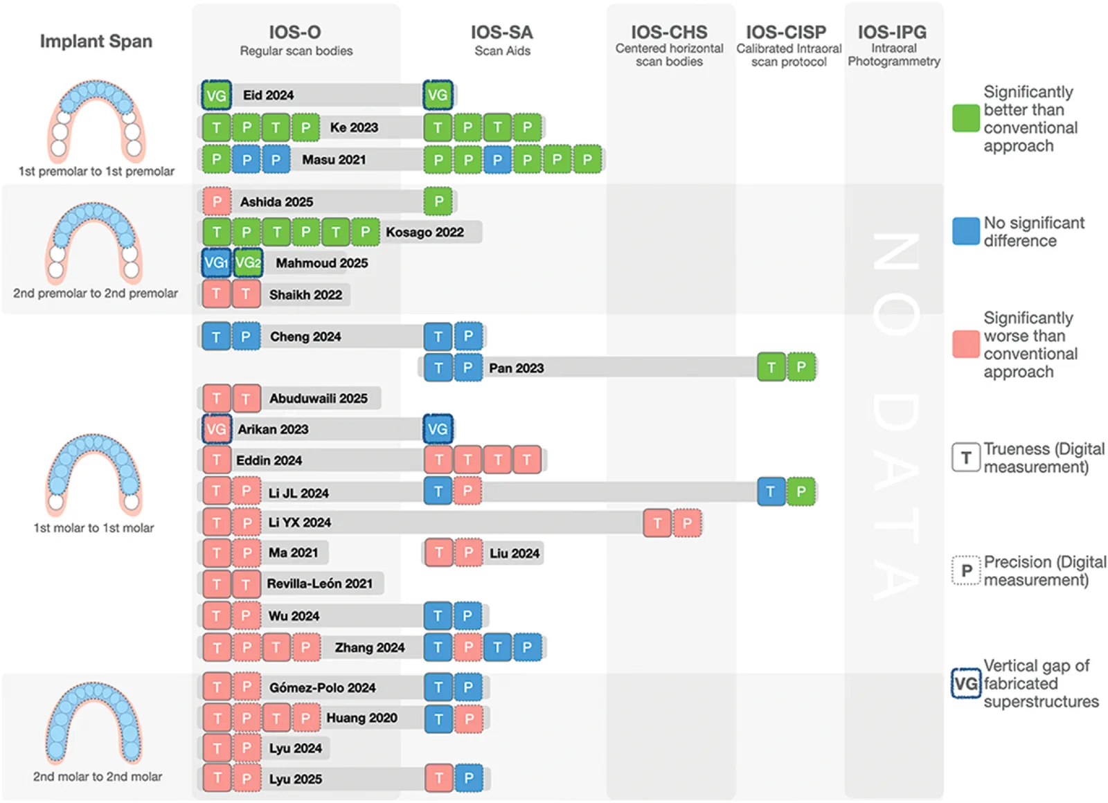
One of the most important findings of the article is that the number of implants is not the only thing that matters; the distance between the terminal implants, or the implant span, also plays a decisive role.
For example, when we have four implants within the range from the right premolar to the left premolar, a regular digital scan in laboratory studies has often shown accuracy comparable to, or even better than, a conventional impression.
But when the implants extend to the molar region on both sides, the situation changes. At these long spans, the conventional splinted impression has generally given more accurate results.
So the longer the implant span, the greater our concern about the cumulative scanning error.
What Can We Do to Increase Scanning Accuracy?
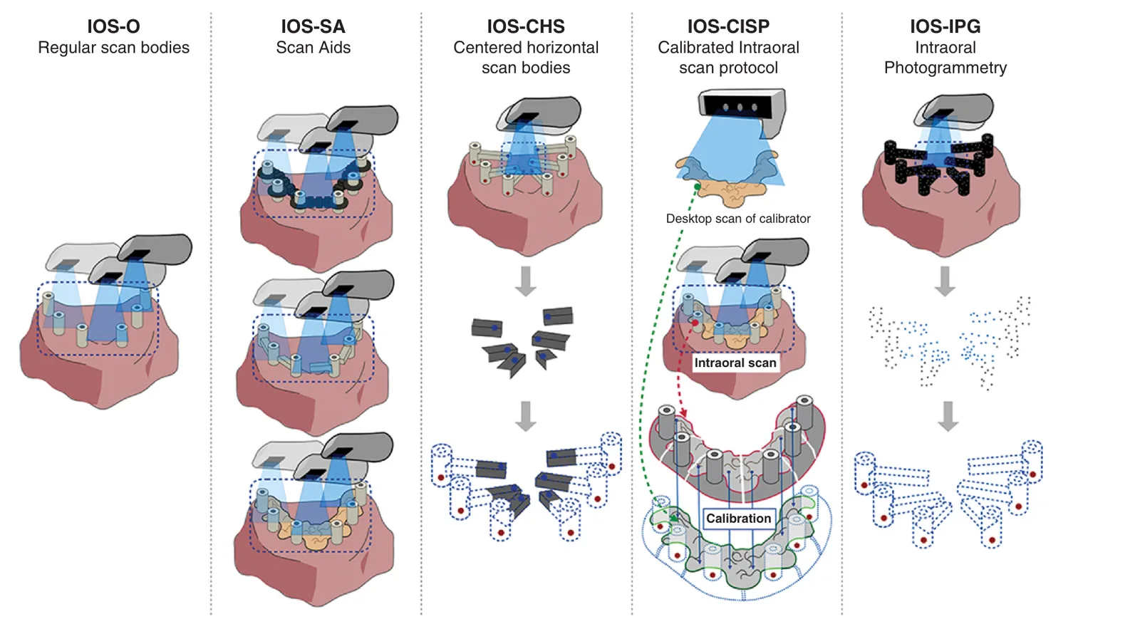
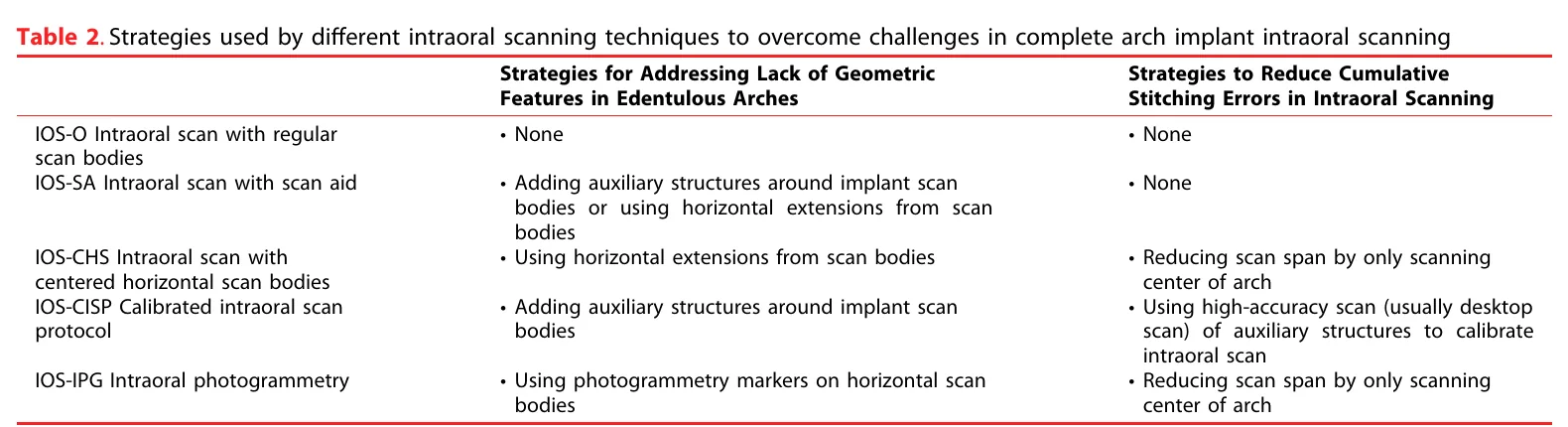
One method is to use a scan aid. These components create more surfaces and geometric landmarks around the scan bodies.
The logic is similar to scanning an arch with natural teeth. When we have more landmarks, the software can join the images more easily, and probably less error occurs.
But the point is that the scan still depends on joining images in sequence. So a scan aid can reduce error, but it does not guarantee correction of cumulative errors, especially when the implant span is long.
Another method is to use horizontal scan bodies that extend toward the center of the arch. In this method, we try to shorten the scanning path so that cumulative errors are smaller.
But the more interesting method is calibrated intraoral scanning, or CISP.
In this technique, an auxiliary structure is attached to the scan bodies and scanned in the mouth together with them. We then scan the same auxiliary structure outside the mouth with a precise laboratory scanner as well.
Now the software has the laboratory scan data as an independent reference and can use it to correct the errors of the intraoral scan.
So the difference between this method and a regular scan aid is that we do not only try to produce less error; we also try to correct the errors that have been produced.
This method has shown promising results even at long spans, but for now there are only two studies on it.
What About Photogrammetry?
In extraoral photogrammetry, the positions of the markers attached to the implants are recorded simultaneously; therefore the problem of sequential stitching, as in a regular intraoral scan, does not exist.
But the article also examined the newer method of intraoral photogrammetry, which uses special markers during the intraoral scan.
For this method, there was no study that directly compared its accuracy with the conventional splinted impression. The evidence on horizontal scan bodies is also very limited.
What Should We Decide Clinically?
If the implant span is relatively short and the scanning conditions are good, an intraoral scan can be a defensible choice.
But when the implants are at the two ends of a long arch, I do not rely merely on having a good scanner or using a scan aid. The conventional splinted impression is still a dependable option.
Of course, we should also consider an important limitation of the article: of the 23 studies, only one was done on a real patient, and none of them evaluated the fit of the fabricated prosthesis in a real patient.
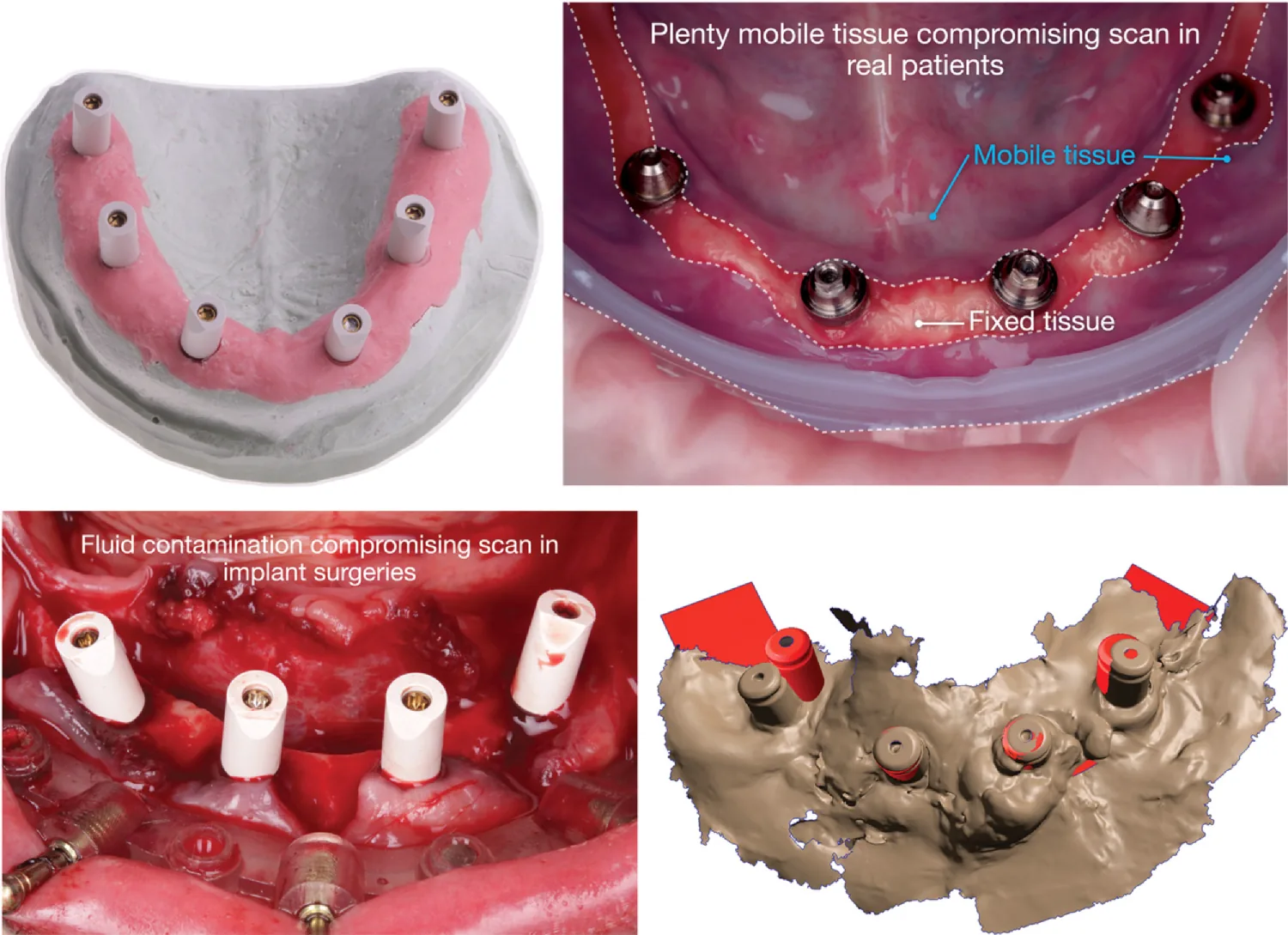
The conditions in the mouth also differ from a laboratory model. Movement of the mucosa and tongue, especially in the mandible, and the presence of blood and fluids during surgery can reduce scanning accuracy.
My Personal Approach
Setting aside the results of this article, in designing fixed full-arch prostheses I prefer not to connect all the implants of the arch with a single monolithic prosthesis.
For example, instead of making a one-piece prosthesis from the right molar to the left molar, I divide it into several independent segments, even if a cantilever results in some areas.
The reason for this decision is not to increase scanning accuracy. The issue is that if there is an error in recording the implant positions, in a long monolithic prosthesis this error may affect the fit of the entire structure. In a multi-segment prosthesis, however, each segment rests on a smaller number of implants and the distance between its supports is shorter. As a result, the effect of cumulative errors can be more limited.
Of course, this approach does not eliminate error, and choosing the cantilever length has its own biomechanical considerations.
This is my personal approach, not a conclusion of the article; but given the problem of cumulative errors that the article raises, its logic can be explained.
Final Word
The message of the article is not that digital scanning is good or bad; the issue is that its accuracy depends on the scanning conditions and, especially, on the distance between the implants.
A scan aid can be helpful, and calibration methods are promising, but there is still not enough clinical evidence to set aside the conventional impression in all conditions.
Our goal is not to digitize treatment at any cost; our goal is to record the implant positions accurately and to make a prosthesis with a proper and dependable fit.