
Chairside 48
When the Abutment Position on the Printed Cast Differs from the Patient’s Mouth
A patient came in for an implant crown. The implant was from a Korean system. Before accepting the case, I checked whether a DIO impression coping was compatible with the implant connection. Once I confirmed compatibility, I scheduled the patient and scanned with a DIO scan body.
When the restoration was ready, I noticed that the crown was about 0.5 mm short. To investigate, I compared the intraoral abutment with the abutment on the printed cast, using the marginal ridge of the first molar as the height reference. The intraoral abutment was about 0.5 mm shorter than the abutment on the cast—the same discrepancy seen in the crown height.
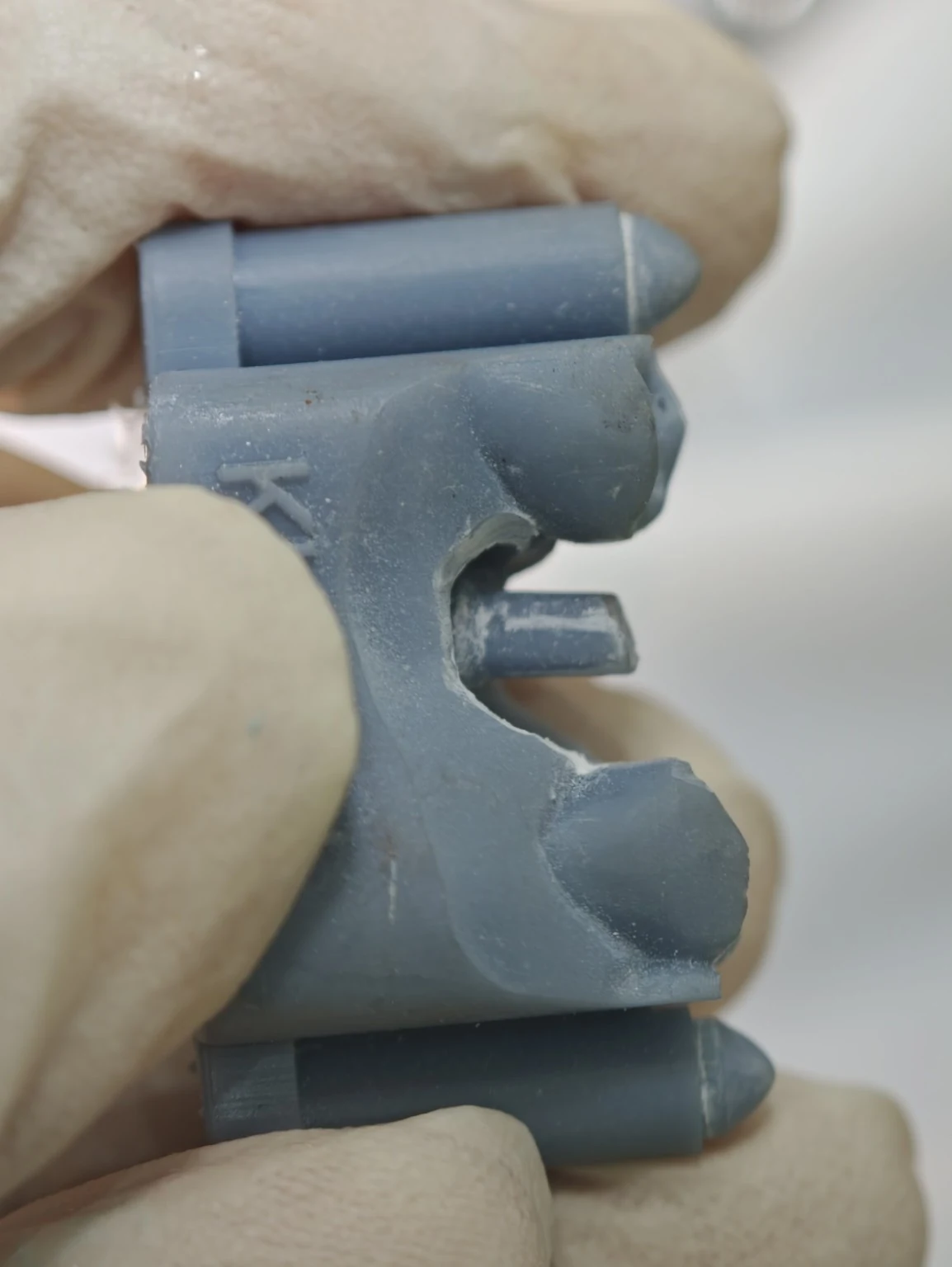
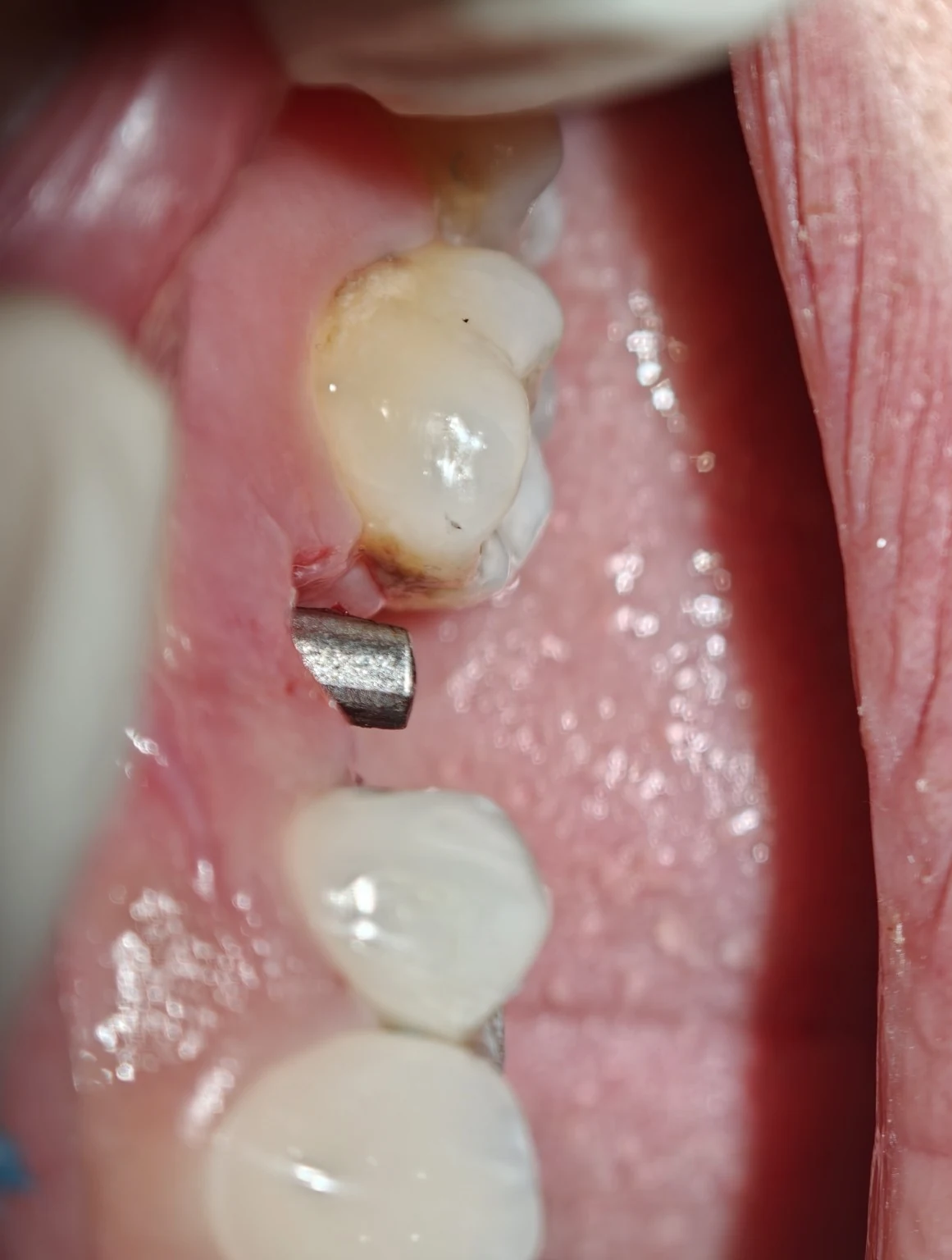
This vertical discrepancy made me suspect that the scan body might not have been fully seated. If the scan body had remained slightly above its final position during scanning, the fixture position could have been recorded more coronally. The cast and crown would then be made from that same recorded information. The original abutment, however, seats fully in the patient’s mouth and sits about 0.5 mm lower than the abutment on the cast.
In this case, the crown was screw-cement-retained, there was no residual cement, and the vertical discrepancy was small. We therefore did not remake the restoration; the height will be corrected with feldspathic porcelain. If the amount of porcelain needed for the correction were excessive, or if a cement-retained crown created a risk of residual cement, the restoration would need to be remade.
Clinical point: When assessing a short crown, comparing the intraoral abutment with the one on the printed cast against a stable reference, such as the marginal ridge of an adjacent tooth, can help identify the source of the discrepancy.
This page is intended for educational use by dentists and dental students.