
MetaNote 22
Diagnosis from a Photograph: What Helen’s Face Teaches Us
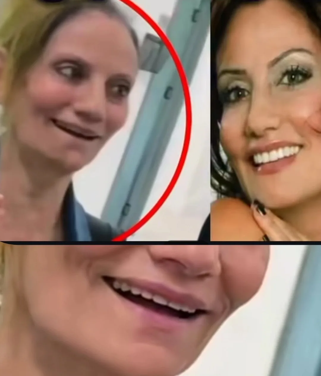
New images of Helen, the veteran singer, have been circulating recently, and the public reaction has been “how much she has aged.” As a prosthodontist, I was tempted to look at these images from a professional angle. The result was instructive for me, but not in the way I expected.
Why Does Helen’s Face Look Aged?
Part of the answer has nothing to do with teeth. Our mental image of Helen belongs to her younger years, and we have not seen her face in all this time. Now we place today’s image next to that mental image, and the gap looks large. We did not see time pass; we see all of it at once.
But the other part of the answer, at least at first glance, was prosthodontic.
The Signs Visible in the Photograph
In the close-up photograph, three signs stood out.
Display of the maxillary anterior teeth during smiling was low; the front teeth showed less than usual.
The smile line was reversed; instead of following the curvature of the lower lip, the incisal edges of the front teeth ran against it.
The lips lacked adequate support from behind, and taken together these pointed to a loss of facial vertical dimension.
What Is Facial Vertical Dimension and Why Does It Age the Face?
Vertical dimension is the distance from the nose to the chin when the teeth are brought together. This distance is determined by the teeth. When teeth are lost, or a prosthesis is made shorter than it should be, this dimension decreases. The mandible then rotates further forward and upward on closing, the lower third of the face becomes shorter and the chin looks more prominent; a condition we call pseudo Class III. What happens is exactly what people call “aging.”
My Guess About the Type of Prosthesis
In one of the images there was no tooth behind the second premolar. Since a denture usually extends beyond the premolars, I guessed that the reconstruction had been done on implants using the shortened dental arch concept; that is, the molars were not replaced and the arch was kept to the premolars. This concept is accepted in its own right and does not by itself reduce vertical dimension.
The likely problem lay elsewhere: when teeth are lost, bone resorbs too. A fixed implant-supported prosthesis has nothing to compensate for this lost tissue, and the lip behind it is left unsupported. So the question belongs to the treatment-planning stage, not to the fabrication of the fixed prosthesis: if resorption was severe enough that the lip needed a flange for support, the right choice may have been a different type of prosthesis from the outset, one that has a flange; a prosthesis that restores lip support and, with a correct vertical dimension and occlusal plane, improves facial form.
A coherent line of reasoning that fit all the visual evidence.
Why Is Diagnosis from a Photograph Unreliable?
Then I watched a few videos of Helen. Some of my assumptions did not match what could be seen in motion.
The reason is clear. A photograph hides several things from us. The lens angle alters facial proportions. Lighting deepens or flattens the shadow beneath the lip and chin. A single frame may have been captured in the middle of a lip movement. And most importantly, a photograph shows neither the occlusion nor the movement of the jaw. Vertical dimension and the state of the lips must be assessed in motion and from several angles, not in a single frozen moment.
The Lesson for the Clinic
The same thing happens in the clinic every day. A photograph patients send of themselves, or even our own clinical photograph, is useful for forming a hypothesis but not sufficient for a treatment plan. Reasoning from an image trains the mind, but the examination makes the diagnosis.
A photograph is good for building a hypothesis, not for a treatment decision; the examination makes the diagnosis, not the image.
The content of this page is prepared for the educational use of dentists and dental students.